Olympus FV3000 Confocal Microscope Will Advance Imaging at UH
On the third floor of the University of Houston’s Science and Research 2 building, research assistant professor of biology and biochemistry Kathleen Gajewski patiently prepares the imaging core’s new confocal microscope for use.
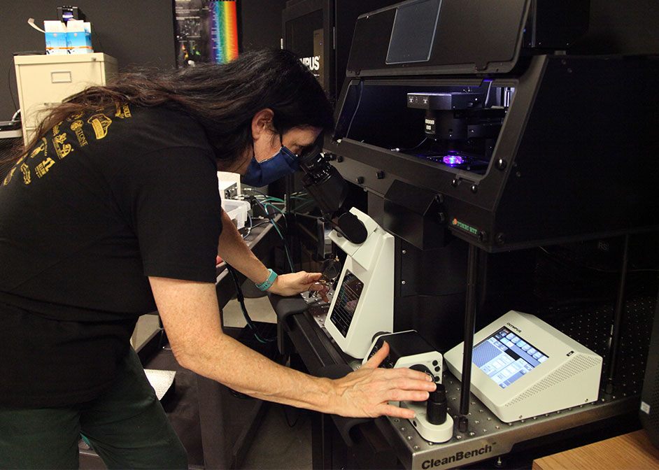
"The problem with most cells is that they’re finicky,” she says. “You have to give them the right environment. To be able to really zoom in on them, you need a microscope in the inverted configuration.”
Thanks to an award from the National Institutes of Health, the UH Biology and Biochemistry Imaging Core, or BBIC, now has a microscope with that configuration. The NIH funding for an Olympus FV3000 laser-scanning inverted confocal microscope will serve the university’s research programs, including several NIH-funded projects that use the Biology and Biochemistry Imaging Core.
The new microscope is housed at the BBIC, which provides affordable access to advanced microscopy instrumentation and provides training to use the instrumentation.
Gajewski is manager of the imaging core, and Arne Lekven, associate professor of biology and biochemistry, is imaging core director and principal investigator on the award.
“Microscopy and imaging technologies are advancing quickly,” Lekven said. “People are making discoveries about how cells and organisms work at a rapid pace. It’s important that our research community has access to the newest technologies available.”
The microscope’s features include live stage imaging, multiple laser lines, spectral detection, and extended depth of focus, critical to the NIH-funded research at UH.
Live cell imaging, in particular, will be of great use, Lekven points out. For example, his lab will use the microscope to study embryonic development.
“We will be able to take time-lapse movies of developing zebrafish embryos,” he said.
The confocal microscope will also be especially useful for labs that study cancer biology, he adds.
“Cancer cells require a specific temperature and specific oxygen and carbon dioxide concentrations in the chamber where the cells are held. Those features are part of this new system.”
A Plethora of Options
Gajewski outlines other key aspects of the microscope, such as a Z-drift compensation feature that checks and adjusts the focus of the microscope and programmable software to track cell migration and to plot vectors.
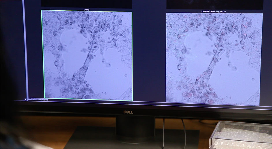
Researchers can use the microscope’s laser scanning technology to take detailed photos of a specimen at fast and slow speeds.
“The old microscope couldn’t switch between both without needing a system reboot,” she said.
The new technology also allows for several different stains – up to 16 – so multiple cell features can be fluorescing at the same time.
“It’s very convenient, because depending on your experiment, you might want to look at the nucleus, at the mitochondria, at the membrane, proteins in the vesicles, especially in the pathology specimens,” said Gajewski.
And while the microscope will certainly be useful for small specimens, Gajewski said it is also beneficial to capture the “big picture,” such as a mouse brain or kidney.
“The microscope has the ability to take several images and stitch them together,” she adds. “You might look at the kidney with a low power objective and make a map, then zoom in on certain areas and take detailed pictures.”
Open to Unique Experiments
For Lekven, receiving the microscope is just the first step for the Biology and Biochemistry Imaging Core.
“My objective for the BBIC is to build on this,” he said, “to keep adding as many new imaging technologies as we can.”
The Olympus FV3000 is open to students of all majors.
“We like unusual requests,” Gajewski said. “If you think this microscope could be of use, we encourage people to come in and talk to us.”
- Rebeca Trejo, College of Natural Sciences and Mathematics