Shoujun Xu and Yuhong Wang Awarded NSF Grant
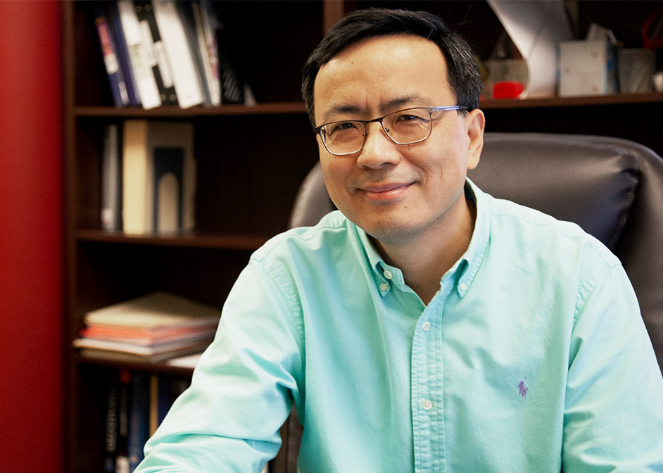
Professor of chemistry Shoujun Xu and professor of biology and biochemistry Yuhong Wang are bringing their expertise together to develop a new imaging technique at the University of Houston.
The researchers are creating a technique called force-modulated fluorescent resonance energy transfer (fmFRET) microscopy. It combines the super-resolution force spectroscopy technique invented by Xu’s lab with the FRET microscopy developed in Wang’s lab.
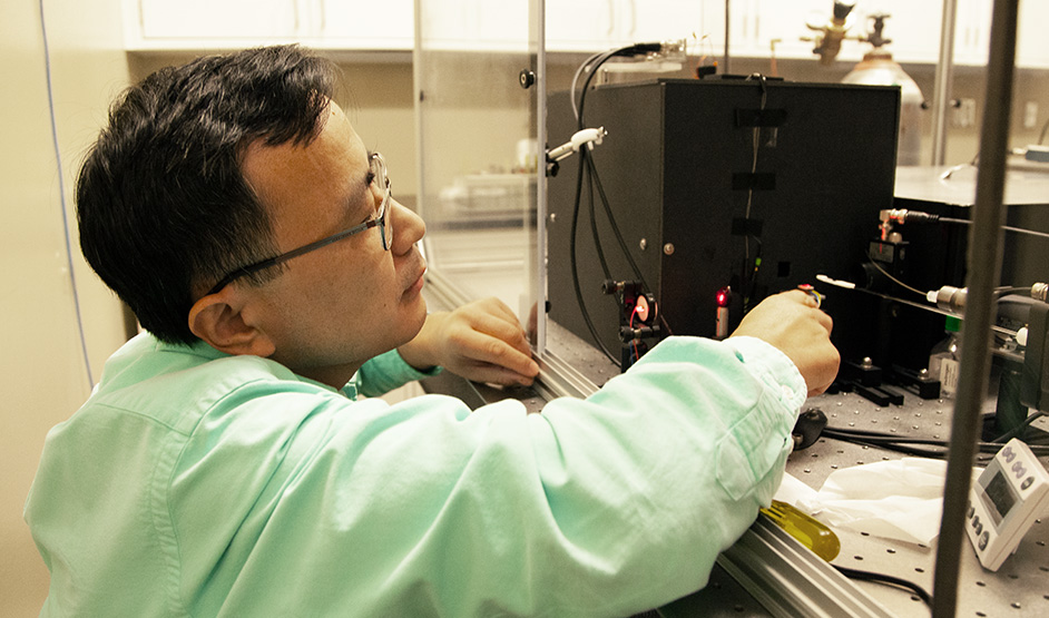
Xu’s force spectroscopy measures the binding strength of two biological entities. Those could be DNA-DNA bonds or protein-protein bonds, for example. With his atomic magnetometer, magnetic particles and acoustic radiation generate a mechanical force that pulls apart the bond between two molecules in order to measure the bond. The precise measurements are at a resolution of about one piconewton. Such a resolution is nearly 10 times better than current competing techniques, sufficient to distinguish DNA-DNA bonds at the smallest unit.
However, “there’s a limitation,” said Xu, principal investigator and faculty at the College of Natural Sciences and Mathematics. “We can only do one sample at a time.”
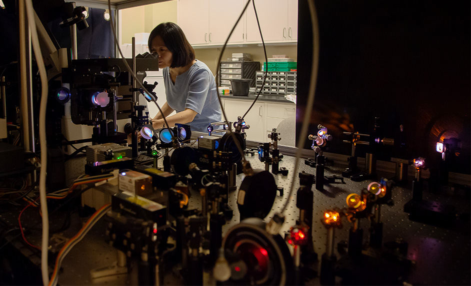
This is where Wang’s microscope comes in to label multiple molecular samples using her fluorescent microscopy.
“The multiplex FRET microscope can detect six pairs of different fluorescently labeled molecules to probe their dynamics,” said Wang.
Paving the Road to Wide Applicability of New Techniques
Two major applications will be explored using the new technique. One application will allow researchers to better understand how the ribosome reads messenger RNA to produce correct proteins and could potentially guide drug design to treat viral infections.
Normally, the ribosome, which makes proteins along the messenger RNA, moves by three nucleotides at a time. However, sometimes, it moves by only two.
“That’s what we call frameshifting,” Xu said. “A lot of viruses use frameshifting to make their different proteins. Our role is to figure out when this happens and how many steps they move.”
Another application of their technique involves widespread use of Xu’s atomic magnetometer in biological research.
“Very, very few chemists, use it for studying biological systems,” he said. “Hopefully we can use what we learned about the force and magnetic detection and expand it with FRET detection to make it much more feasible to other researchers.”
Both Xu and Wang thank UH’s Division of Research for providing the equipment necessary for their experiments. “Developing this new technique would not have been possible without DOR’s support,” Xu said.
Their work is funded by a recent $519,045 grant from the National Science Foundation. Xu and Wang have collaborated for more than 10 years.
- Rebeca Trejo, College of Natural Sciences and Mathematics