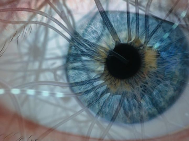

In the high-speed supercomputer that is the human brain, where quick connections are made in response to outside stimuli, neurons (or individual cells) race to transmit information using electrical signals. Also cranking in the brain’s motherboard, electrical synapses, or gap junctions, transmit information through the direct flow of electrical current at those junctions.
Gap junctions are made of protein channels that physically connect adjacent cells, assisting in the rapid exchange of small molecules and ions and playing an essential role in a wide range of physiological processes in nearly every system in the body, including the nervous system.
Though powerful, gap junctions are small, and studying them is difficult, so they tend to be under-reported or simply ignored. Now, two studies at the University of Houston are taking close looks at gap junctions.
“A greater understanding may guide the development of new treatments or diagnostics for brain disorders and degenerative diseases, but because of their small size, gap junctions are not visible in large-scale serial Electron Microscopy (EM) data sets,” said neuroscientist Christophe Pierre Ribelayga, professor of optometry at the UH College of Optometry, who has received a $1.8 million grant from the National Institute of Mental Health to provide a way to analyze gap junctions in great detail.
“The main innovation of this work is the development of an approach that will break some of the major roadblocks that have hindered our understanding of fundamental integrative processes supported by electrical synapses,” said Ribelayga.
To develop an approach to image gap junction connectivity in EM datasets, Ribelayga keeps a keen eye on how the neural retina (in the back of the eye) processes information during day and night and the role of gap junctions in that process. Specifically, he will study regions of the retina that contain gap junctions of dramatically different sizes and shapes, to correlate structure and function. His technical approaches can be readily applied to other brain regions, too.
Gap junctions are having quite a moment. The transfer to UH of a separate investigation, titled Regulation of Retinal Gap Junctions, funded with $1.2 million from the NIH’s National Eye Institute, has been announced by the project’s principal investigator John O’Brien, professor of optometry at the UH College of Optometry. The project will continue its 20-year investigation of gap-junction plasticity, which affects not just the retina but also a wide range of other neurologic functions.
“Gap junctions in the retina profoundly influence how the retina extracts and processes a visual scene, and they have the ability to reconfigure neural circuits to adapt to dim or bright conditions” O’Brien said. “This project has made more new advances in understanding the underlying molecular mechanisms than others like it. We’ve made a lot of progress.”
The next goal is to work out how the signal pathways reach deep into cell frameworks. In the longer view, O’Brien hopes to explore common links that may bring solutions to problems as common as myopia and as rare as certain childhood seizure disorders.
Interestingly, no gap in knowledge exists between the two researchers. Ribelayga and O’Brien often work together, and each intends to continue the quest to uncover the secrets that lie within the junction gaps.